Research
A major theme of my research is developing both software and hardware for high-throughput computational 3D imaging techniques. My interests span the broadest, most inclusive interpretation of “3D”, ranging from dense 3D tomography to sparse 3D topography, from the microscale to the mesoscale and macroscale. Some projects include:
- High-speed 4D fluorescence light field tomography of whole freely moving organisms
- High-throughput computational 3D imaging with camera arrays
- Optical coherence refraction tomography (OCRT)
- Multiview imaging over extremely wide angular ranges
- Diffraction tomography with a deep image prior
High-speed 4D fluorescence light field tomography of whole freely moving organisms
We developed a new technique, Reflective Fourier Light field Computed Tompography (ReFLeCT), which can capture synchronized video from perspectives spanning 2π steradians across 10s of cubic mm volumes of view, enabling multimodal volumetric video at up to 120 fps. While existing volumetric imaging approaches applied to model organisms such as zebrafish and Drosophila larvae require immobilization, ReFLeCT’s high spatiotemporal throughput enables 4D imaging of whole organisms as they are freely moving!
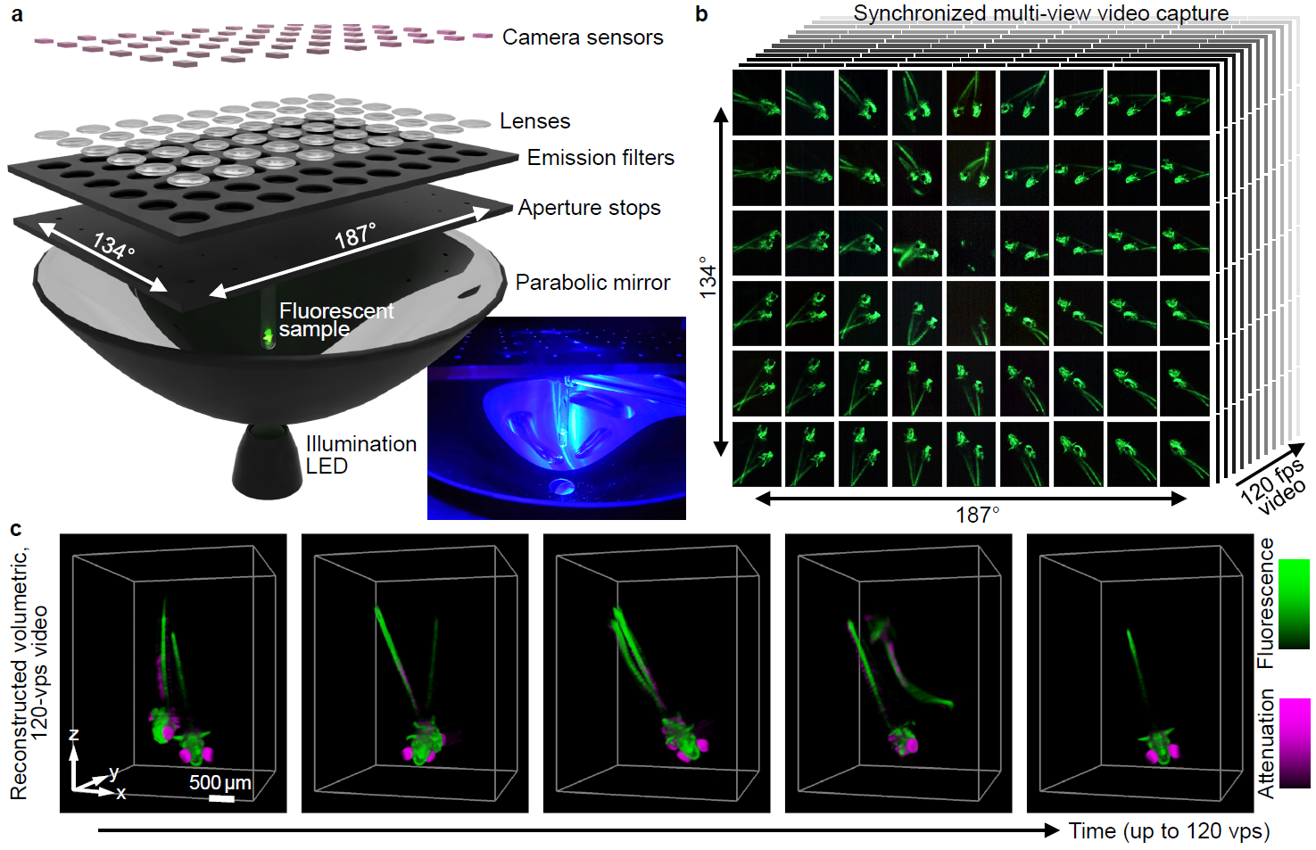
Demonstration of ReFLeCT applied to a freely moving Drosophila larva expressing GFP in its pericardial cells:
ReFLeCT applied to a freely moving zebrafish larva:
The left 6×9 panels of the above two videos are the synchronized raw multi-view video capture, and the right 2×2 panels are four different visualizations of the 4D reconstruction.
Relevant publications
- KC Zhou et al., High-speed 4D fluorescence light field tomography of whole freely moving organisms, bioRxiv 2024.09.16.609432 (2024)
- KC Zhou et al., High-speed multi-view imaging approaching 4pi steradians using conic section mirrors: theoretical and practical considerations, JOSA A 38, 1810-1822 (2021)
Code
- Visualization: https://github.com/kevinczhou/ReFLeCT-4D-visualization
- 4D reconstruction: coming soon!
High-throughput computational 3D imaging with camera arrays
We present 3D-RAPID (3D Reconstruction with an Array-based Parallelized Imaging Device), a computational 3D imaging technique based on an array of 12-megapixel cameras capable of imaging at extremely high spatiotemporal throughts (>5 gigapixels/sec). 3D-RAPID features a spatiotemporally scalable, physics/self-supervised algorithm that enables simultaneous 3D reconstruction and stitching of arbitrarily many cameras across arbitrarily many temporal frames.
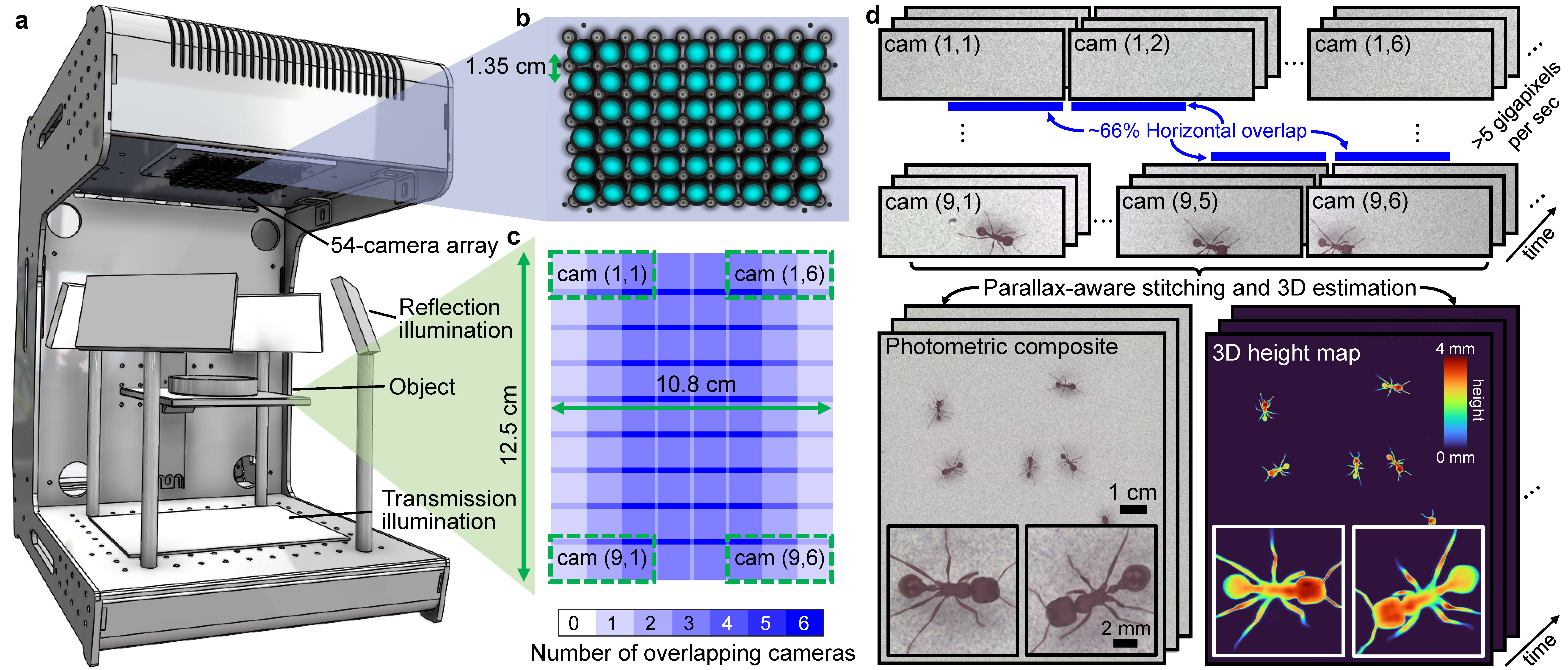
Our high spatiotemporal throughputs (>5 GP/sec) enable high-resolution 3D imaging of freely behaving organisms over wide FOVs (>130 cm2) at high speed (up to 230 fps):
The left panel shows the zoomed-out view of the entire arena of freely swimming zebrafish; the right panels show the zoomed-in individually tracked zebrafish. The video alternates between photometric (RGB) and 3D height information.
Freely moving harvester ants (left: photometric frame, right: 3D height map).
For more videos, see the supplementary materials of the accompanying publication below. See also https://gigazoom.rc.duke.edu/ for interactive views of full video frames.
Relevant publications
- KC Zhou et al., Parallelized computational 3D video microscopy of freely moving organisms at multiple gigapixels per second, Nature Photonics 17:442–450 (2023)
- KC Zhou et al., Computational 3D topographic microscopy from terabytes of data per sample, Journal of Big Data 11(62) (2024)
- KC Zhou et al., Mesoscopic photogrammetry with an unstabilized phone camera, CVPR (2021)
Code
- https://github.com/kevinczhou/3D-RAPID
- https://github.com/kevinczhou/starcam
- https://github.com/kevinczhou/mesoscopic-photogrammetry
Optical coherence refraction tomography (OCRT)
OCRT is a new computational 3D microscopy technique that incorporates extreme angular diversity to incoherently enhance resolution and reduce speckle of conventional OCT.
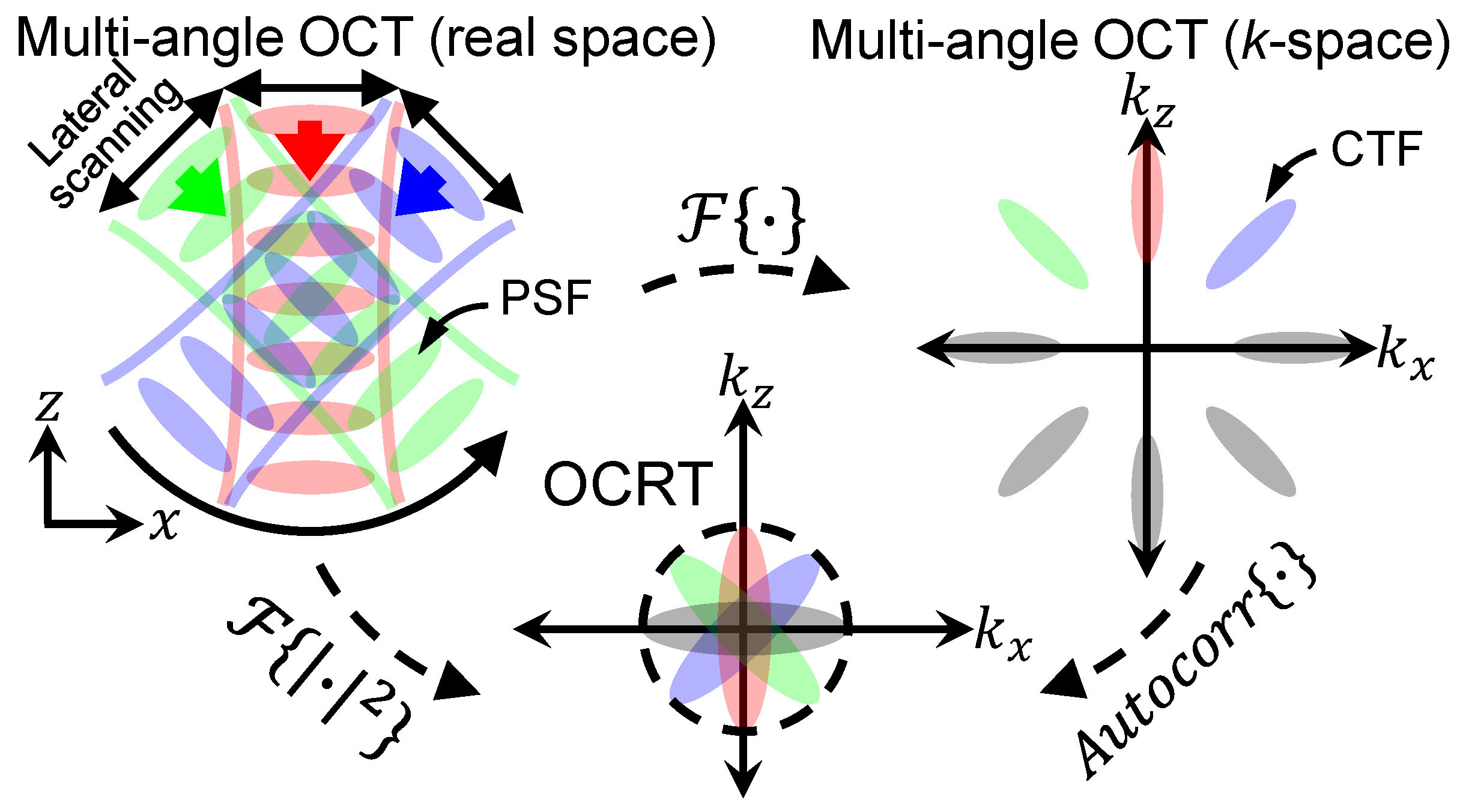
OCRT registers multi-angle OCT images or volumes by solving an inverse refraction problem, a process that also computationally reconstructs a 3D refractive index map of the sample. The following videos demonstrate the marked improvement of 3D OCRT over conventional OCT volumetric data (zebrafish larva and mouse esophagus, respectively):
Relevant publications
- KC Zhou et al., Computational 3D microscopy with optical coherence refraction tomography, Optica 9, 593-601 (2022)
- KC Zhou et al., Unified k-space theory of optical coherence tomography, Advances in Optics and Photonics 13, 462-514 (2021)
- KC Zhou et al., Spectroscopic optical coherence refraction tomography, Optics Letters 45, 2091-2094 (2020)
- KC Zhou et al., Optical coherence refraction tomography, Nature Photonics 13, 794–802 (2019)
Code
- https://github.com/kevinczhou/3d-ocrt
- https://github.com/kevinczhou/optical-coherence-refraction-tomography
Multiview imaging over extremely wide angular ranges
We introduce a class of techniques that can achieve multi-view imaging over extremely wide angular ranges (theoretically up to 4pi steradians) based on conic section mirrors (e.g., parabolic or ellipsoidal mirrors). Although traditionally such mirrors are known to exhibit off-axis aberrations and are thus more widely used for on-axis focusing or collimation, we show theoretically and practically that they are particularly well-suited for multi-view 3D imaging applications.
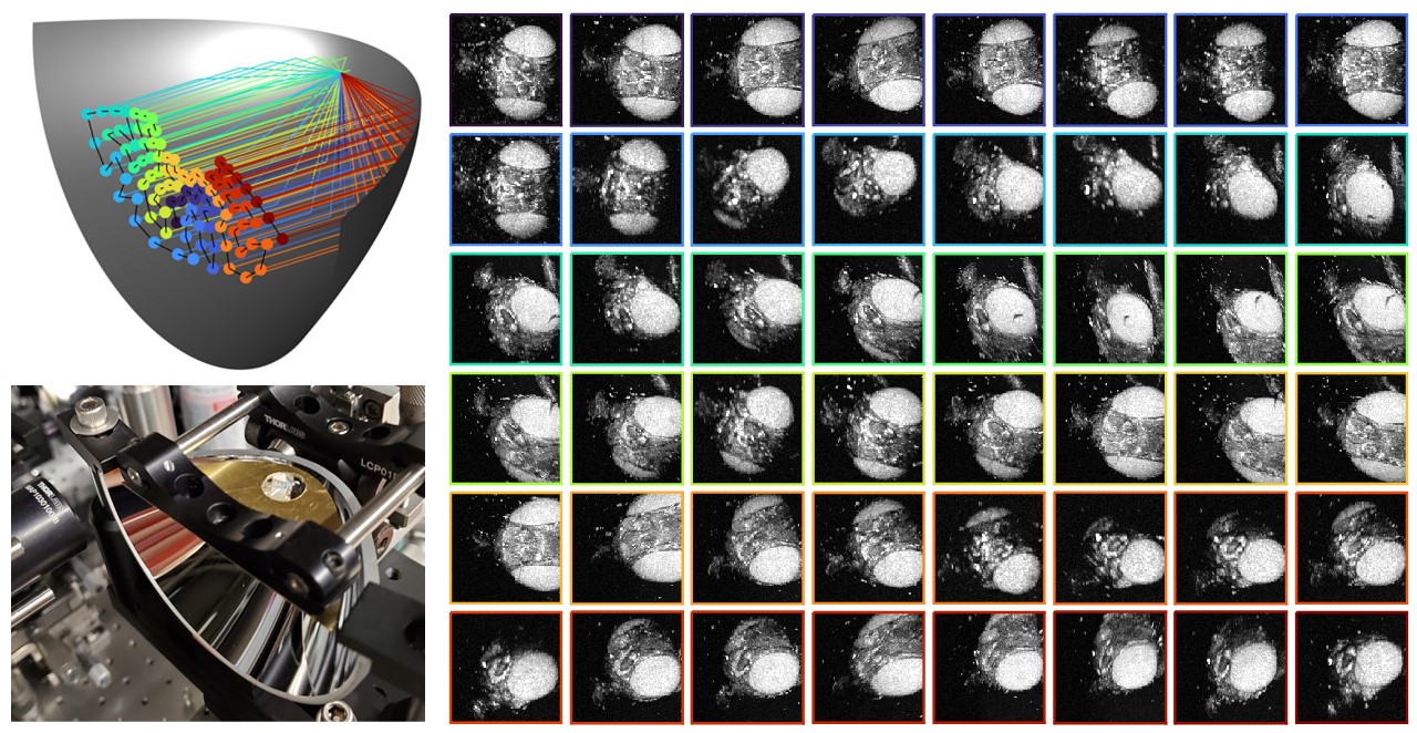
The above figure shows multi-view OCT imaging of a fruit fly head from up to ±75°. The fruit fly images are color-coded by view angle, as denoted by the ray traces on the left. These images were used for 3D OCRT reconstructions.
Relevant publications
- KC Zhou et al., Computational 3D microscopy with optical coherence refraction tomography, Optica 9, 593-601 (2022)
- KC Zhou et al., High-speed multi-view imaging approaching 4pi steradians using conic section mirrors: theoretical and practical considerations, JOSA A 38, 1810-1822 (2021)
Diffraction tomography with a deep image prior
Optical diffraction tomography is a coherent 3D imaging modality that reconstructs a 3D refractive index (RI) distribution based on multi-angle diffraction patterns. A well-known problem particularly when using low-NA objectives is the “missing cone” centered about the kz axis in the Fourier domain, which reduces the axial resolution. At the same time, however, low-NA objectives tend to have higher space-bandwidth products (SBPs), a property exploited in Fourier ptychography for 2D samples.
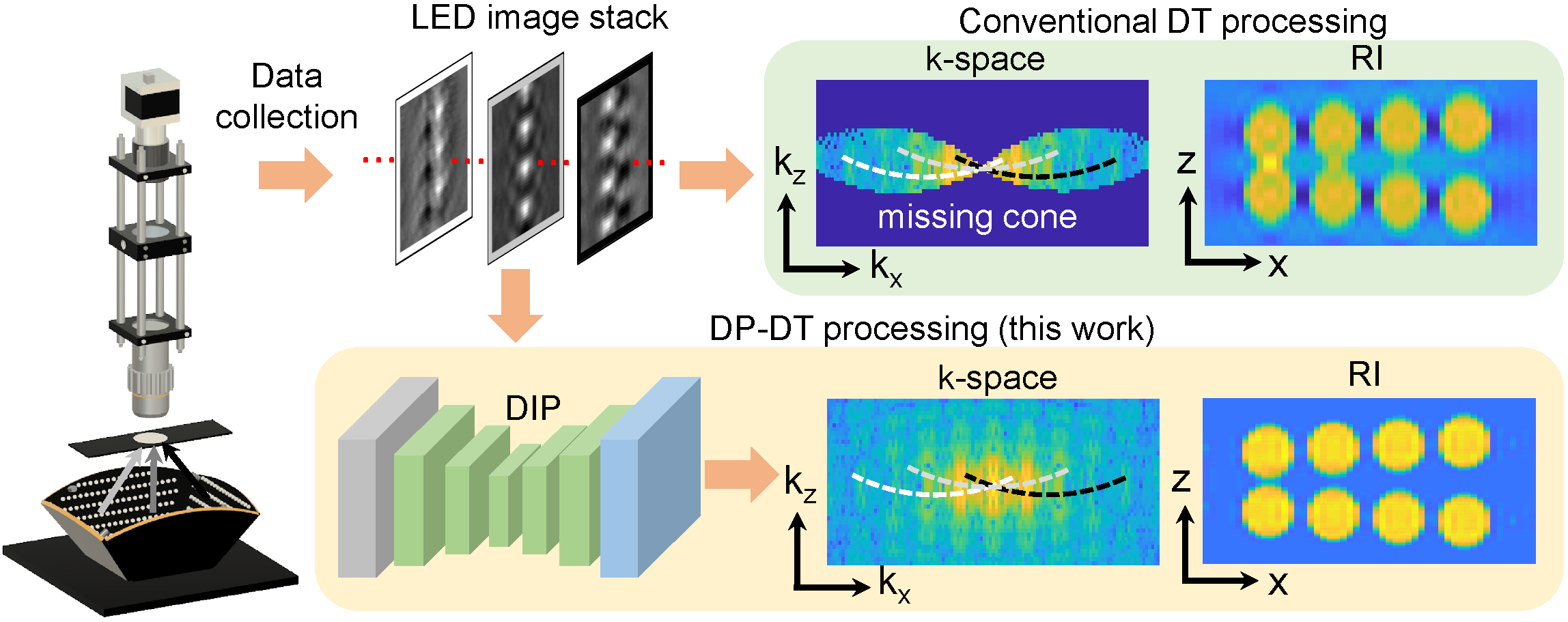
To combat the missing cone problem, we reparameterized the 3D RI distribution as the output of an untrained CNN, and optimized its parameters instead of the 3D volume directly during the 3D reconstruction process. Interestingly, the inherent biases of the CNN structure, even without pretraining, are enough to fill in the missing cone!
Relevant publications
- KC Zhou & R Horstmeyer, Diffraction tomography with a deep image prior, Optics Express 28, 12872-12896 (2020)
Code
